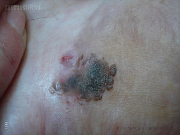
Меланоакантома: вариант себорейного кератоза
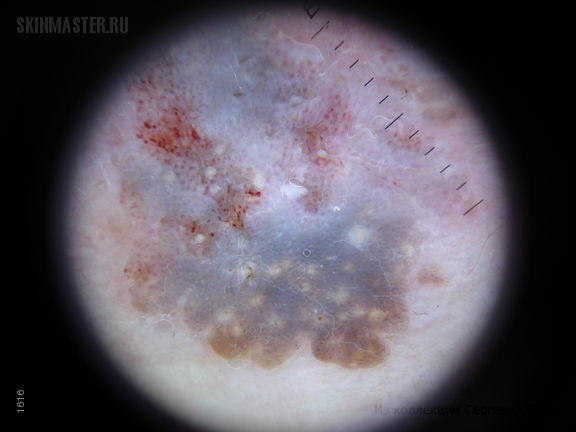
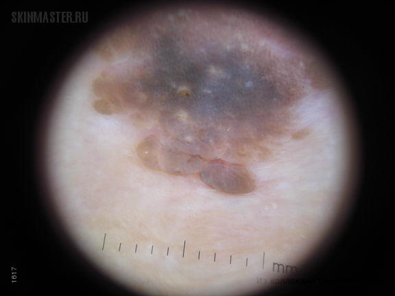
При дерматоскопическом исследовании определяется структура себорейного кератоза с роговыми пробками и псевдокистами. В местах травмирования вследствие натирания и мацерации определяются точечные геморрагии. Также можно выявить мелкоглыбчатую диффузную пигментацию.
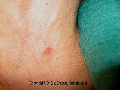
A 62-year-old woman consulted for this cervical lesion which was present for 10 months.
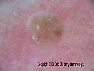
Dermoscopy
revealed sharp demarcations, scales and some hairpin-like vessels.
The lesion was excised and pathology revealed a seborrheic keratosis.
The main clue was the presence of hairpin-like vessels.
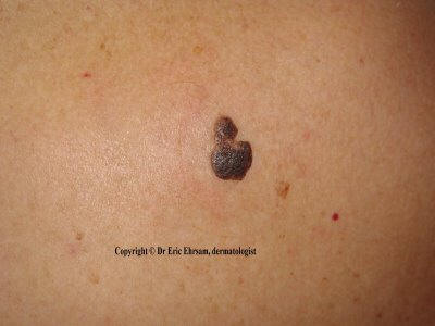
A 50-year-old woman consulted for an enlarging and pruritic lesion on her back.
 This pigmented
lesion was characterized in dermoscopy by:
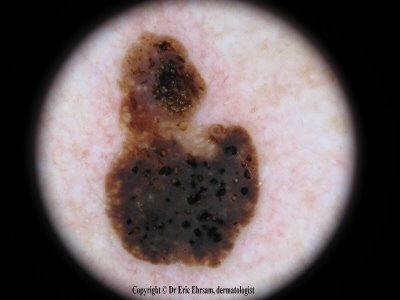
This pigmented
lesion was characterized in dermoscopy by:absence of pigment network
sharp demarcation
presence of milia-like cysts
comedo-like openings
This dermoscopic presentation was typical of a seborrheic keratosis.