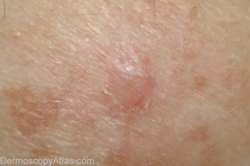
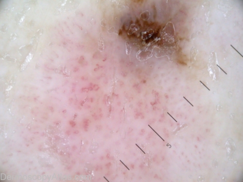
History: 60 year old woman with long history of BCCs, new lesion upper arm since last check 6 months prior. Lesion today is superior of two, inferior is another BCC. Histology reported as; Sections confirm a level 1 superficial spreading malignant melanoma arising in a dysplastic naevus. There is no ulceration and no invasion is seen.
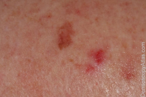
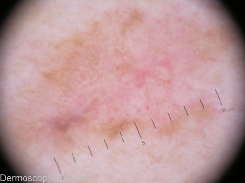
Description: Manipulated image to emphasise vascular structure. Dot vessels predominate
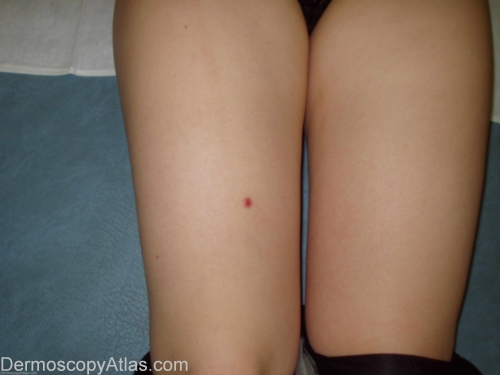
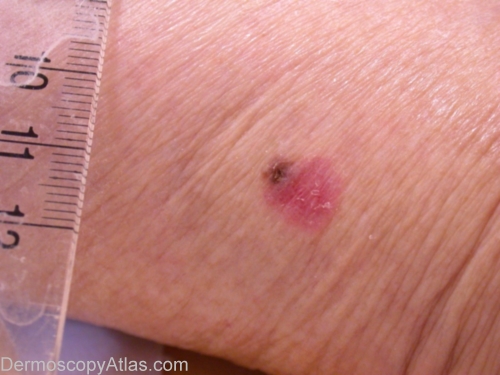
History: From words of
the patient this lesion of skin of the leg has appeared 2 years ago and
gradually increased in size. Last year is marked by an itch in a zone around
the lesion. The pathology showed an amelanotic melanoma: Breslow 1.75,
Clark II
Sex: F
Age: 21
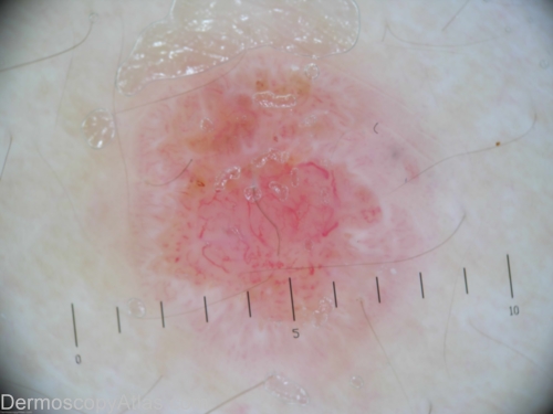
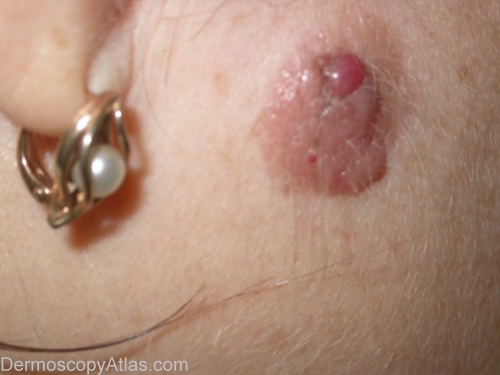
Description: An irregular heavily vascularised lesion. In the central part you can see arborizing vessels and on the periphery hairpin vessels.
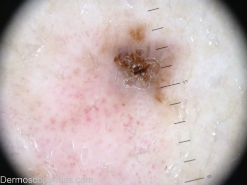
History: This lesion was discovered by a systematic exam. Clinically it was not much concerning, but as the dermoscopic view showed no clear (benign) sign, excision became mandatory. The pathology revealed a compound melanocytic proliferation, with a strong and atypical junctional activity. The final diagnosis being Melanoma in situ, which developed from a compound nevus.
Sex: M
Age: 70

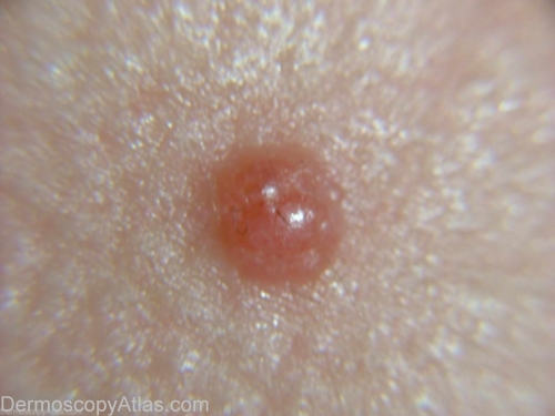
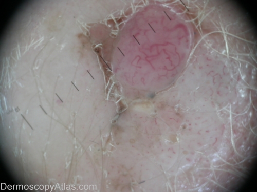
Description: The lesion shows mostly vessels, which fit neither with a diagnosis of seborrheic keratosis, nor with a basal cell carcinoma: mainly dotted vessels, and irregular linear vessels. There also white areas of regression, and an inversed network. It is circled by a brown edge, but it is difficult to find any sign in favour of a network. Finally, a blue-grey clod was a pitfall, in favour a basal-cell carcinoma.
Description: The clinical view: a "Bowen-like" lesion, with a brown crust at one pole.
History: A 69 years old lady who came for this chronic lesion of the inferior part of the calf. No precise chronology. It was a pink, scaly, diameter 1 cm plaque, with a pigmented crust at his superior pole. Dermoscopy showed a polymorphous vascular pattern, with some corkscrew vessels, and for the pigmented pole, mainly a crust which was surrounded by blue-grey pigment.The pathology revealed an invasive melanoma, SSM, Clark IV, Breslow 1.7.
Clue to diagnosis: Polymorphous vessels in association with areas of blue-grey pigment.
Description: The crusted area surrounded by grey-blue pigmentation. Below, the polymorphous vessels.
Description: Close-up on the vessels: polymorphous vessels, with hairpin vessels, corkscrew vessels, some irregular coma vessels.
History: A 43-year-old woman consulted for a pigmented lesion on her cheek. The lesion was present from birth. She noticed some changes 1 or 2 years ago (see pink lesion).
Dermoscopic description: Comma vessels, dotted vessels in the periphery and some corkscrew vessels occurring in a pink lesion.
Description: Dermatoscopic description: Comma vessels, dotted vessels in the periphery and some corkscrew vessels occurring in a pink lesion.
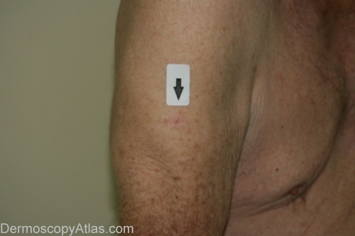
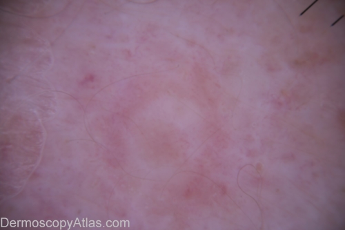
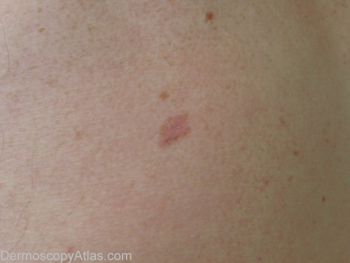
History: This 87 year old veteran of World War 2 had a regular skin examination ( Past history of non-melanoma skin cancers) and this small papule could not be confidently diagnosed clinically. It was thought to be benign but NMSC needed exclusion. It came back after a 4mm punch biopsy removal of the visible lesion as a level 3 amelanotic melanoma (Breslow thickness 1.3mm) with complete regression of the epidermal component. A further 1.5 cm clearance was obtained and the report came back as superficial spreading amelanotic melanoma (small residual level 2 0.3 mm thick)cleared by only 5mm. In retrospect, especially with enhancement of the image by Dr. Lester Cowell, the large insitu amelanotic melanoma can be visualised.