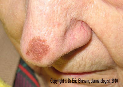
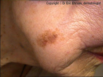
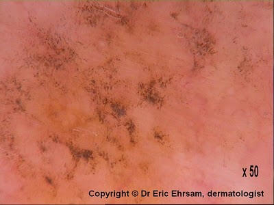
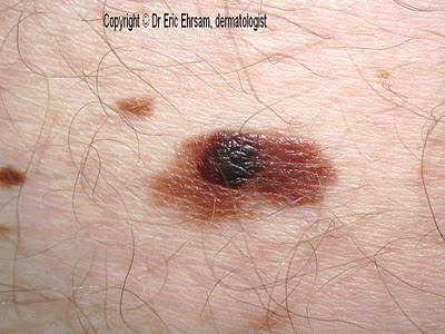
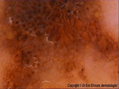
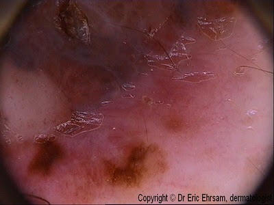
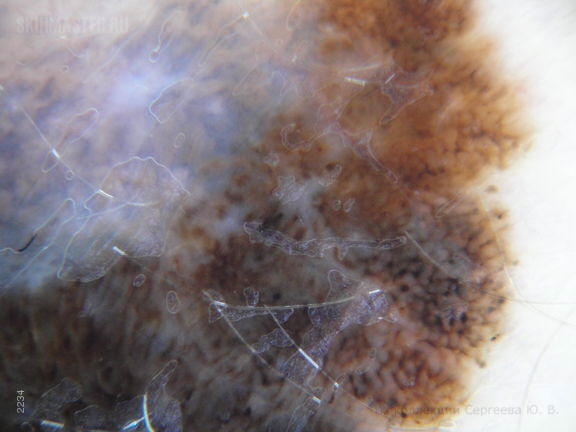
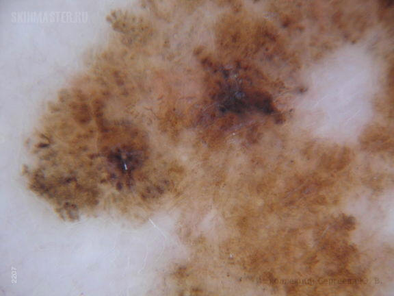
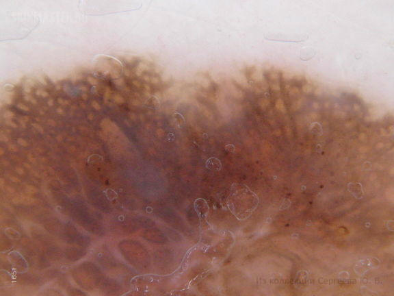
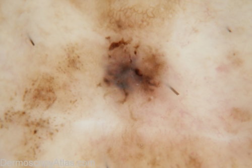
A 85-year-old woman consulted for a pigmented lesion on the tip of her nose.
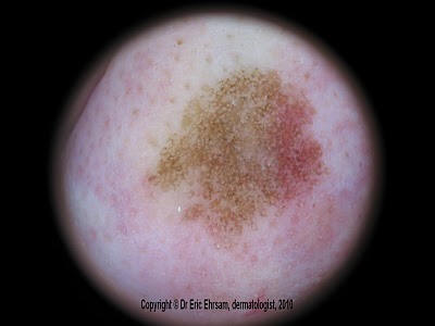
 Dermoscopy revealed:
a pseudo-pigmented network (due to the facial localisation)
asymmetric pigmentation of the follicular openings
slate gray dots disposed as annular- granular structures
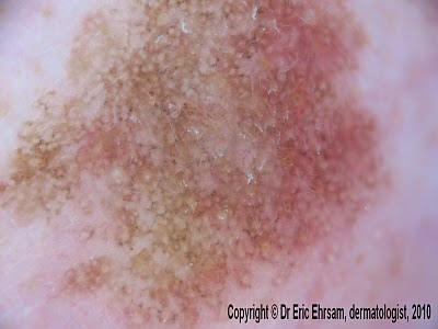
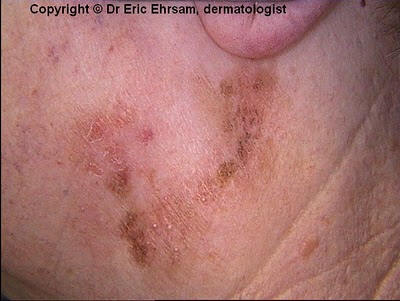
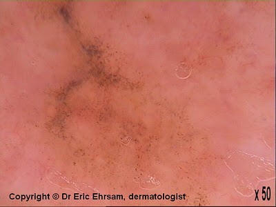
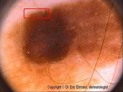
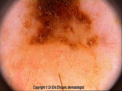
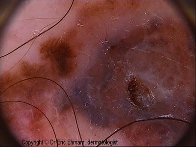
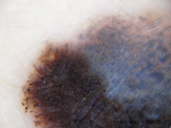
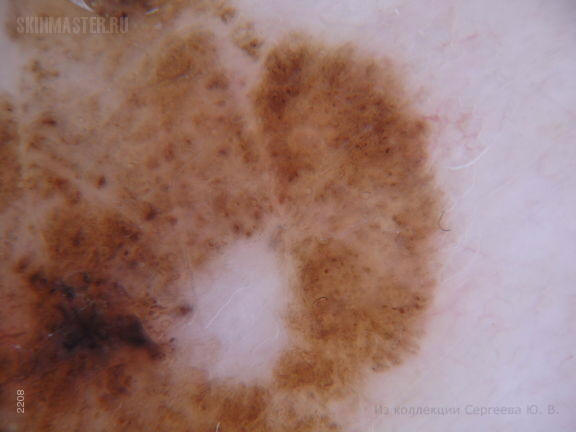
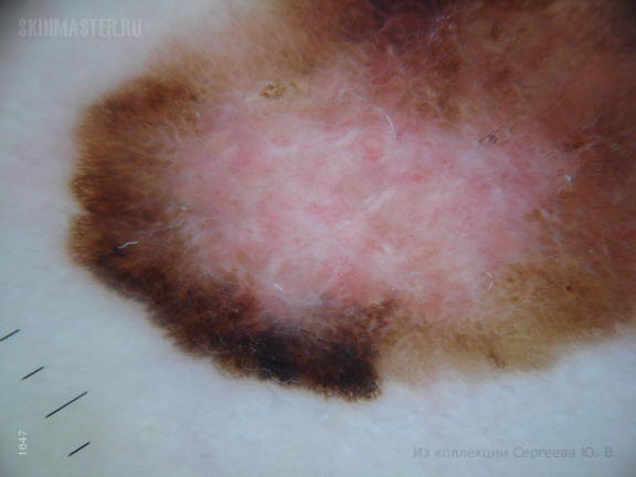
A 85-year-old woman consulted for a slowly enlarging pigmented lesion on her right cheek
 Dermoscopy revealed a
pseudonetwork which is characteristic of facial pigmented lesions.
Dermoscopy revealed a
pseudonetwork which is characteristic of facial pigmented lesions.Furthermore, dermoscopy showed multiple slate-grays dots or pepper-like granules realizing a typical an annular-granular pattern or peppering in favor of a lentigo maligna melanoma. A biopsy confirmed this diagnosis.
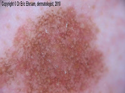
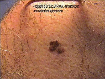
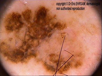
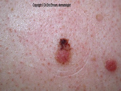
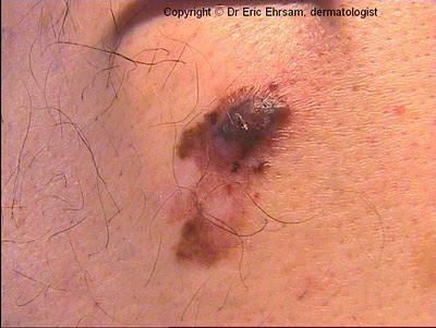
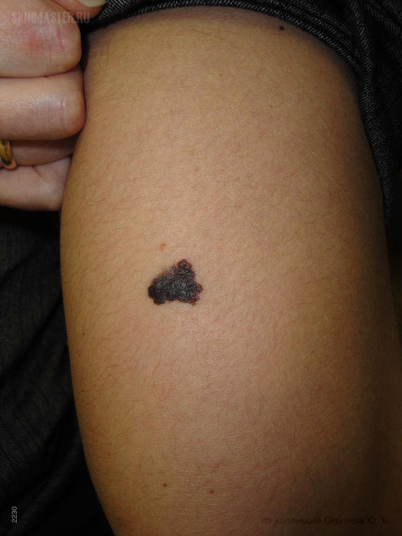
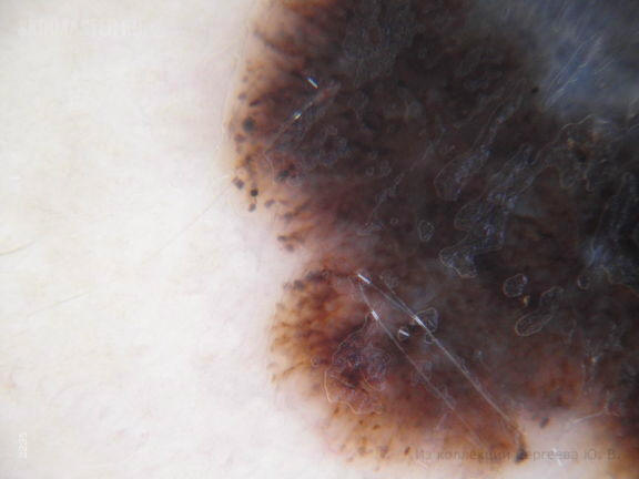
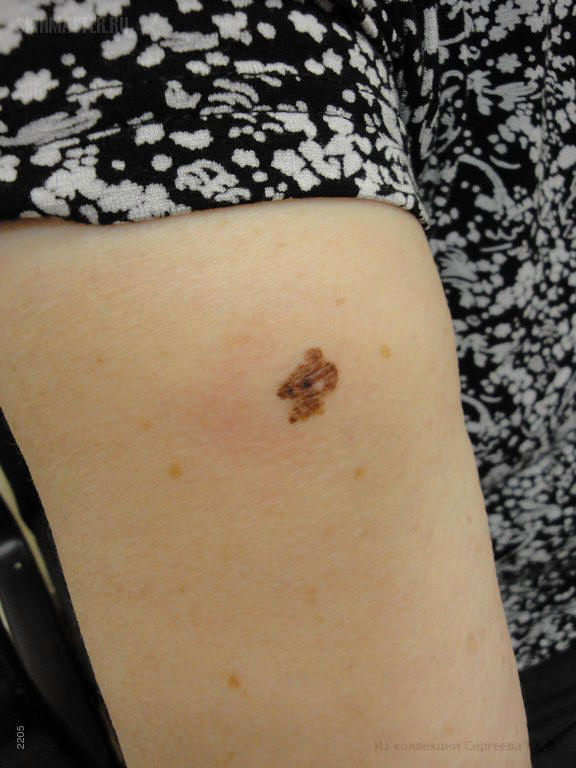
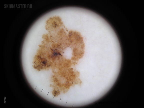
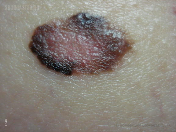
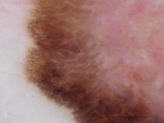
A 70-year-old woman consulted for this light-brown pigmented lesion on her left cervical area.
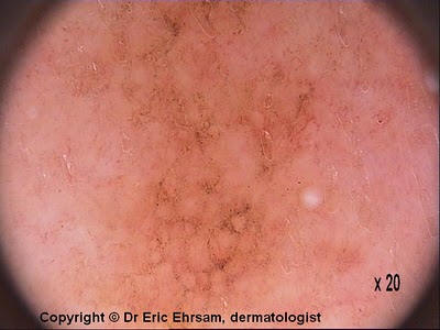
Dermoscopy revealed slate-gray dots disposed in an annular-granular pattern in favor of a lentigo maligna melanoma which was confirmed by biopsies performed on the lesion.
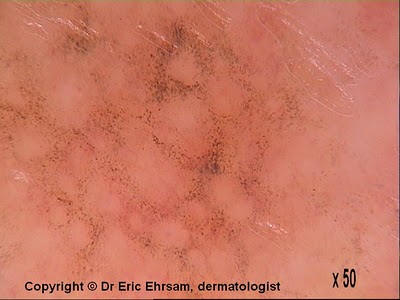
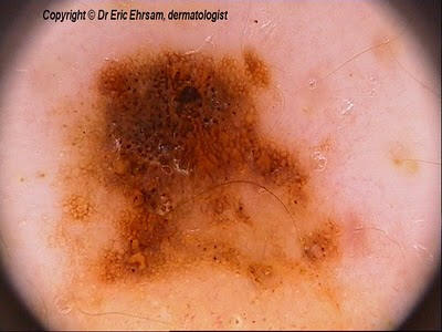
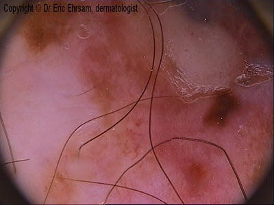
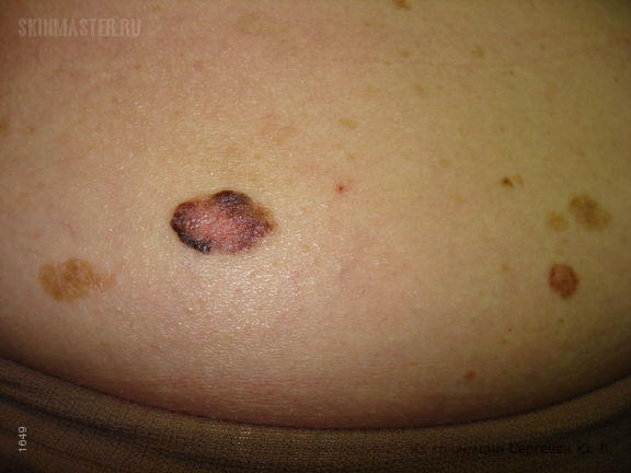
A 62-year-old patient with a superficial spreading melanoma on his right shoulder.
Dermoscopy (x20) revealed white areas (scar-like depigmentation) and delicate peppering.
A 50-year-old man presented an irregularly pigmented lesion his back.
Dermoscopy revealed an asymetric pigmentation, multiple colors, streaks (red circle), and a delicate blue white veil in favor of a superficial spreading melanoma. The lesion was removed with a 2 mm margin and histology confirmed the diagnosis. The Breslow index was measured 0.63 mm. A re-excision was performed with a 1 cm margin. No mestastases were detected.
A 60-year-old man consulted for a changing of color of one of his nevi on his back. The upper part of this nevus turned dark brown a few months before.
On the upper part of the lesion, dermoscopy revealed an atypical reticular pattern, asymmetric irregular globules and a blotch.

On the lower part of the lesion, there was an homogeneous non pigmented pattern with some comma vessels in favor of a dermal nevus. The tumor was excised and pathology revealed a superficial spreading melanoma developed on a pre-existing dermal nevus.
A 62-year-old man consulted for this multicolored tumor on his scapular area. This lesion was clinically in favor of an advanced invasive melanoma (nodular melanoma).
 Asymmetrical
pigmentation pattern, multiple colours , atypical vessels, atypical
reticular pattern, blue-white veil, and ulceration were found in this
case of invasive melanoma, Breslow thickness 9mm
Asymmetrical
pigmentation pattern, multiple colours , atypical vessels, atypical
reticular pattern, blue-white veil, and ulceration were found in this
case of invasive melanoma, Breslow thickness 9mm
 При
дерматоскопическом исследовании выявляется классическая
дерматоскопическая картина меланомы, с такими признаками как
бело-голубая вуаль,атипичная пигментная сетка, краевое распределение
пигментных глыбок, регрессирующие структуры и др.
При
дерматоскопическом исследовании выявляется классическая
дерматоскопическая картина меланомы, с такими признаками как
бело-голубая вуаль,атипичная пигментная сетка, краевое распределение
пигментных глыбок, регрессирующие структуры и др.
 При
дерматоскопическом исследовании выявлены признаки, характерные для
меланомы: псевдоподы, неправильных очертаний и размеров пигментные
глыбки и пятна, скопление пигментных зерен по периферии очагов и многие
другие признаки.
При
дерматоскопическом исследовании выявлены признаки, характерные для
меланомы: псевдоподы, неправильных очертаний и размеров пигментные
глыбки и пятна, скопление пигментных зерен по периферии очагов и многие
другие признаки.
Lentigo maligna (поверхностно-распространяющаяся меланома)
 Меланома,
поверхностно-распространяющаяся форма
Меланома,
поверхностно-распространяющаяся форма
Во всех случая выявления меланомы или при подозрении на нее, рекомендую принимать меры срочного характера. Несмотря на то, что родинка существует много лет, наиболее грозным симптомом является ее возвышение по медиальному краю, отмечаемое в последние дни, что может свидетельствовать о ее переходе в фазу вертикального роста.
History: This 71 year old lady presented with this macule in continuity with a scar 4 years after excision of a level 4 lentigo maligna melanoma which had been excised with 1.5 mm clearance at the closest margin. The initial exision biopsy of the pigmented macule at the lower end of the scar revealed level 1 lentigo maligna melanoma involving the entirety of both margins. Two further attempts at surgical clearance resulted finally in a level 1 lentigo maligna melanoma with lateral clearance of greater than 10 mm.
Description: Dermoscopy with Dermlite Fluid Pattern - Brown clods, blue-grey structureless area superimposed Colours - brown, blue-grey Clues - blue-grey area,eccentric focal brown dots, polymorphous vessels