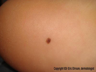
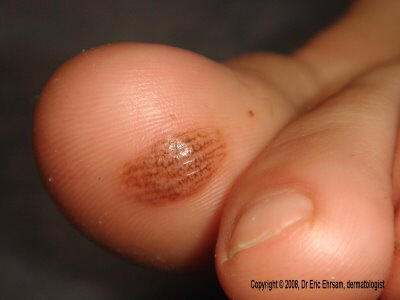
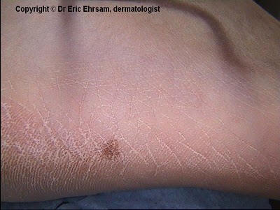
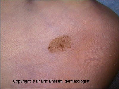
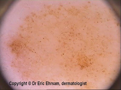
A 14-year-old girl consulted for this small plantar nevus.
Dermoscopy revealed a typical lattice-like pattern which is a subtype of parallel furrow pattern in favour of a benign acral plantar nevus.
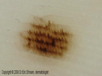
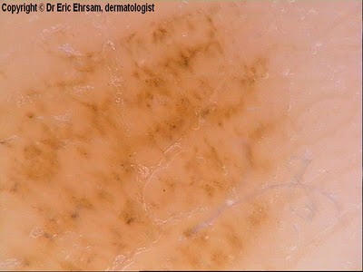
A 48-year-old woman consulted for this pigmented lesion on her 5th right finger present since early childhood.
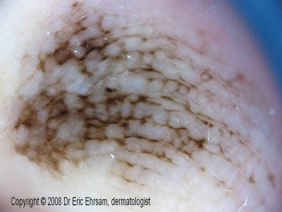
Dermoscopy revealed a parallel furrow pattern and in some areas a lattice-like pattern.
In parallel furrow pattern, the pigmentation follows the sulci at the
difference of parallel ridge pattern where the pigmentation follows the
cristae.
In Acral melanocytic nevi, 3 types of patterns can be observed:
parallel furrow pattern
lattice-like pattern
fibrillar pattern
Parallel ridge pattern are typical of acral melanomas but can be
observed in other cases:
milk-line nevus
subcorneal hemorrhages (talon noir)
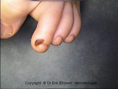
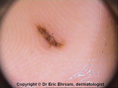
A 17-year-old girl consulted for an acral melanocytic lesion on her left toe.
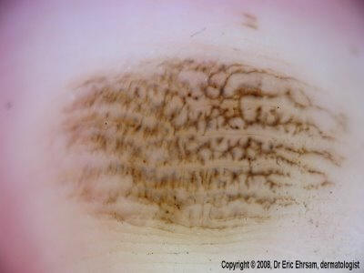 Dermoscopy revealed
a lattice-like pattern in favor of a benign acral melanocytic nevus.
Dermoscopy revealed
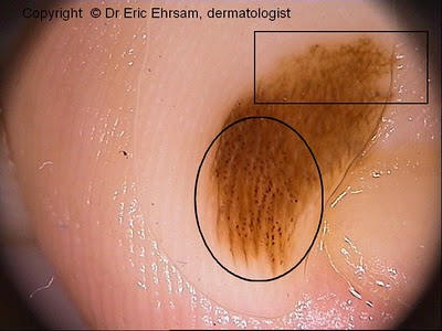
a lattice-like pattern in favor of a benign acral melanocytic nevus.Lattice-like pattern is a subtype of parallel furrow pattern.
It is more often localized on arch areas at the difference of fibrillar pattern which is observed more frequently on pressure areas.
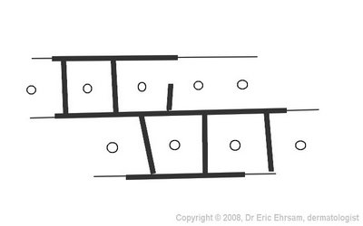
On this figure above, the lattice-like pattern corresponds to the longitudinal and transversal thicker lines and the white circles symbolize eccrine pores.
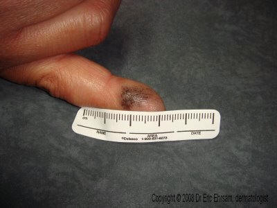
A 48-year-old woman noticed a pigmented lesion on the right side of her right foot.
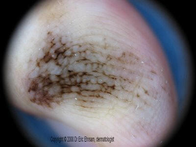
 Dermoscopy revealed
a fibrillar pattern in favor of a benign acral melanocytic nevus.
Dermoscopy revealed
a fibrillar pattern in favor of a benign acral melanocytic nevus.
Fibrillar pattern is a variant of parallel furrow pattern.
A 6-year-old boy consulted for a congenital acral melanocytic nevus on his 2nd left toe.
 This lesion
exhibited a combination of 3 patterns, namely a parallel furrow pattern
(dots on the furrows), an homogeneous pattern and a reticular pattern.
This lesion
exhibited a combination of 3 patterns, namely a parallel furrow pattern
(dots on the furrows), an homogeneous pattern and a reticular pattern.
Parallel furrow pattern (circle) and reticular pattern (rectangle) were the main patterns observed on this benign melanocytic nevus.
A 25-year-old man consulted for a congenital melanocytic nevus located on the right plantar arch.
 Dermoscopy featured
a globular pattern without signs of parallel pattern.
Dermoscopy featured
a globular pattern without signs of parallel pattern.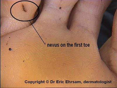
Typical case of benign acral melanocytic nevus.
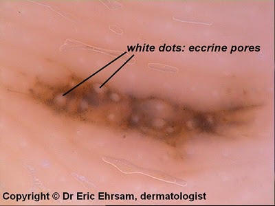
Eccrine pores are well recognized as white dots which are located on the ridges between the furrows